[et_pb_section bb_built=”1″][et_pb_row][et_pb_column type=”4_4″][et_pb_text _builder_version=”3.13.1″]
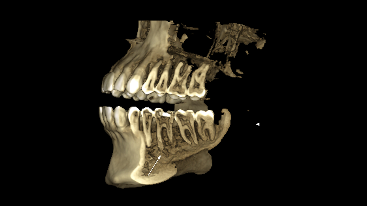
X-rays of the bones of the body use a very small dose of ionizing radiation to produce images of each bone and is usually used to determine fractures, tumors, or conditions that cause wear (degeneration) of the bone.
Structures that are dense (like bone) will block most of the x-ray particles. These areas will appear white. The metal and special dye used to highlight areas of the body will also appear white. The muscle, fat and liquids will appear as gray shadows and the segments containing air will appear black. Currently a team of researchers from New Zealand has developed the first radiography that can transmit images with flat or curved faces, and with full-color depth using a device based on the original X-ray machine. This device uses a particle detection technology called Medipix, this technology acts as a subatomic camera that provides a high contrast image. Color x-rays clearly show the differences between muscles, bones, and cartilage, as well as the size and location of cancerous tumors. Definitely a great technological breakthrough in medicine.
These and other innovations are now possible in Pharmamedic.
[/et_pb_text][/et_pb_column][/et_pb_row][/et_pb_section]