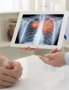
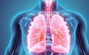
When pulmonary edema occurs in a short period of time, it is usually called acute pulmonary edema and, in this case, it is a medical emergency, since the patient’s respiratory capacity may be compromised.
The lungs fill with fluid when blood that is normally inside the blood vessels leaks out and floods the alveoli of the lungs. Of course, this only happens when something is wrong with the lungs or the heart itself; Thus, we can divide pulmonary edema into the following types:
- Cardiogenic pulmonary edema
- Non-cardiogenic pulmonary edema
- Neurogenic pulmonary edema
All pulmonary edemas have in common that they cause a feeling of shortness of breath, which is also known as dyspnea.
The most common cause of pulmonary edema is usually congestive heart failure, which in turn can be caused by a sudden rise in blood pressure, aortic valves, or heart attacks or heart problems.
However, pulmonary edema can also be due to other causes, such as taking certain drugs, kidney failure, lung damage caused by poisoning or infection, being exposed to high altitudes.
cardiogenic pulmonary edema
The heart receives blood from the entire body in the right atrium and from there it passes to the right ventricle, which drives it to the lungs to oxygenate the blood. Afterwards, all the blood from the lungs is collected in the left atrium, and passes through the mitral valve to the left ventricle, where it is pumped out to the rest of the human body. Any disease that causes the left side of the heart to malfunction will cause blood to back up into the lungs, where it pools. This malfunction is known as left heart failure (the left side of the heart is not efficient enough to pump all the blood it needs). Situations in which left heart failure occurs are:
- Acute myocardial infarction: when part of the heart muscle is destroyed, the heart loses pumping power.
- Cardiac arrhythmias: the heart loses the proper rhythm of pumping and blood is not ejected properly.
- Mitral stenosis: A narrowing of the mitral valve would make it more difficult for blood to pass from the left atrium to the left ventricle.
- Mitral rupture: if the mitral valve ruptures, the blood that should leave the left ventricle towards the aorta would return to the left atrium and the lungs.
- Hypertensive crisis: a sudden increase in systemic blood pressure makes it very difficult for the left ventricle to expel blood to the rest of the body.
- Chronic heart disease: the heart is an organ that is affected by many diseases, which do not always have a devastating effect on it, but do deteriorate it little by little. These sick hearts are usually compensated by natural mechanisms or drugs indicated by the doctor, but situations that require a greater functioning of the cardiac pump can cause heart failure. The situations that most frequently produce this effect are physical exercise, increased blood pressure, lack of oxygen in the environment, infection, bleeding, anemia, pregnancy, and hyperthyroidism.
- Fluid overload: it is common for patients who need it to be hydrated intravenously in the hospital. An excess of fluid through this route can make it impossible for the heart to mobilize it, and heart failure then occurs.
Non-cardiogenic Pulmonary Edema:
This includes the causes that cause an invasion of the lungs by fluid, but without the heart being diseased. In this case, the problem arises in the walls of the blood capillaries that supply the lungs, since they lose their ability to retain blood inside and allow fluid to pass into the alveoli. The causes are very varied:
- Aspiration of gastric juices: in unconscious patients the cough reflex is diminished and this means that, if they vomit, the gastric juices and all the contents that are in the stomach rise to the larynx and go down to the lungs through the windpipe. Acids cause damage to the alveolar walls, which allow fluid to pass through them.
- Sepsis: Septic patients are those who have such a degree of infection in their body that it is possible to find bacteria or toxins in their blood.
- Trauma: In contusions of the lung there is direct rupture of the blood vessels, but not only can edema of the lung be produced in this way, it is also possible that trauma anywhere in the human body triggers an exaggerated inflammatory response (as in sepsis), and substances reach the lung that increase the permeability of the capillaries.
- Pneumonia: localized infection in the lungs can have a direct effect on the surrounding capillaries.
- Major burns: People who suffer major burns to their skin develop systemic inflammation that causes lung edema by the same mechanism as sepsis.
- Acute pancreatitis: in an inflammation of the pancreas part of this organ is destroyed; thus, the enzymes and acids that are inside are released into the arteries and cause damage to the human body in a generalized way. Specifically in the lung, it damages the blood capillaries and allows liquid to flow into the alveoli.
- Inhalation of toxic gases: certain chemical substances have a direct harmful effect on the pulmonary alveoli and capillaries.
- Misuse of drugs: there are drugs that can cause alterations in the pulmonary capillaries directly or indirectly, either by overdose or by taking medicines that are not indicated.
- Drowning: the simplest cause of the lungs filling up with liquid is sucking in water from the outside, which is what happens in drowning people.
Neurogenic Pulmonary Edema:
This is the least common form of pulmonary edema. It happens in people who have a brain injury from trauma or a tumor, and also in heroin addicts when high doses of drugs are injected. When the brain’s control over the hypothalamus is removed, it stimulates the sympathetic nervous system, which is involuntary and is responsible for internal organ functions, such as increasing blood flow to the lungs during physical exercise. By increasing pulmonary blood flow unnecessarily, excess blood flows out into the alveoli, flooding them.
The symptom common to all types of pulmonary edema is the feeling of shortness of breath, which in medicine is known as dyspnea. This can occur more or less intensely, and be established more or less quickly. When the pulmonary edema is acute, the dyspnea is very intense and occurs suddenly (a situation that usually occurs in a myocardial infarction, mitral valve rupture, or aspiration of gastric juices). The patient literally drowns due to the flooding of his lungs and it is an emergency situation.
When pulmonary edema develops more slowly, symptoms vary depending on whether the pulmonary edema is cardiac or noncardiac in origin.
Symptoms of cardiogenic pulmonary edema:
The degree of dyspnea corresponds to the degree of heart failure causing it:
- Grade I: the person can perform a physical activity without limitations and without getting tired.
- Grade II: there is a slight limitation of physical activity, but during rest there is no sensation of shortness of breath.
- Grade III: minimal physical activity causes fatigue.
- Grade IV: the patient is unable to perform any physical activity, and even at rest suffers from fatigue and shortness of breath.
Another common symptom in people who have this type of pulmonary edema is the need to sleep with several pillows under the head, to sit up and thus reduce the feeling of shortness of breath. The rest of the symptoms will depend on your own heart failure.
Symptoms of non-cardiogenic pulmonary edema:
Supportive treatment of pulmonary edema
- Oxygen: Oxygen by mask should be administered to all patients with acute pulmonary edema. The flow must be continuous and with a concentration of between 60-90% of oxygen in the inspired air.
- PEEP: is the positive pressure at the end of inspiration. It is a device that transmits a positive pressure to the lungs, which allows the alveoli to distend and favor the exchange of oxygen.
- Mechanical ventilation: only if the previous measures do not work.


