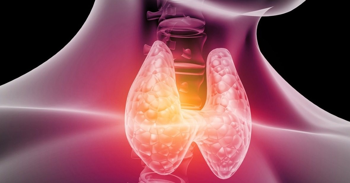
Its function is the synthesis of thyroid hormone, which plays an important role in regulating metabolism.
It is necessary to know a series of concepts related to the increase in the size of the thyroid to understand what it is about.
- Simple goiter: enlargement of the thyroid gland that is not accompanied by hyperthyroidism, hypothyroidism, neoplastic (cancer), inflammatory or autoimmune process. Also called non-toxic or normal-functioning goiter.
- Thyroid nodule: it is any mass of the thyroid with a consistency different from that of the normal gland.
- Toxic nodular goiter: Toxic nodular goiter grows from a simple goiter and occurs more often in older people.
There are also various classifications of goiter based on different parameters:
- Size: goiter is graded from grade 0 (no goiter) to grade 4 (giant goiter).
- Shape: diffuse, nodular or multinodular goiter.
- Epidemiological criteria: endemic goiter (occurs in a certain region where goiter prevalence is relatively high as a result of iodine deficiency), or sporadic (does not occur in a particular population).
- Cause that produces goiter.
- Functionality: functioning or non-functioning goiter.

Goiter treatment will vary depending on the symptoms it produces. In many cases, expectant management is chosen, which consists of monitoring the evolution of the goiter over time. In others, when it causes many symptoms, more aggressive treatments are chosen, such as irradiating the thyroid, or even removing part or all of it.
Although there are many causes that can cause the appearance of goiter, the specific mechanism by which the increase in thyroid size occurs remains unknown. It has been found that most patients have subtle disturbances of hormone formation. This inability of the thyroid to produce or secrete hormones, together with a normal or high level of TSH (a hormone synthesized in the pituitary gland that stimulates the thyroid to form thyroid hormone), would lead to an enlargement of the gland in an attempt to compensate.
The main known causes of goiter are:
- Iodine deficiency: it is the most common cause of goiter.
- Inflammation of the thyroid due to different causes: thyroiditis, infections, radiation.
- Goitrogens (substances that can favor the appearance of goiter): monovalent anions, tobacco, lithium, iodine, sulfonylureas, salicylates, soybean, sunflower, walnut, and peanut oils.
- Autoimmune thyroid disease: Hashimoto’s thyroiditis and Graves-Basedow disease.
- Congenital alterations.
- Infiltrative diseases: Riedel’s thyroiditis, amyloidosis, hemochromatosis.
- Benign and malignant tumors.
- Puberty, pregnancy.
- Other causes: acromegaly, oral contraceptives, hydatidiform mole, etc.
Goiter is more common in women, probably due to the higher prevalence of autoimmune diseases and the increased need for iodine during pregnancy and for estrogen during adolescence.
Finally, it should be emphasized that the thyroid gland increases in size over the years, so that around the eighth decade of life, many people have goiter due to the presence of one or more thyroid nodules in the thyroid gland.
Most patients have no symptoms at the time of diagnosis, and the presence of a goiter is discovered incidentally during a physical examination performed for another reason. On other occasions, the patient goes to his doctor for noticing the appearance of a lump or tumor of variable size on the anterior face of the neck, which may or may not be painful on palpation.
The most frequent complication of goiter, when it is large, is the compression of neighboring structures found in the neck, thus causing symptoms in the patient such as difficulty breathing, irritative cough, difficulty swallowing, hoarseness or changes in the voice. Despite everything, these symptoms are not very frequent.
In patients in whom the goiter is so large that it enters the retrosternal region, raising the arms can cause respiratory distress, dizziness, and even syncope.
The prevention of these complications is based on an early diagnosis and correct medical treatment. If, despite this, compression of neighboring structures occurs, the treatment will be surgical.
To reach the diagnosis of goiter, both the anamnesis (clinical interview conducted by the doctor about the patient’s symptoms) and the physical examination are very important, but numerous imaging tests are available that allow a very good view of the thyroid anatomy to be obtained. , which allows the diagnosis to be made.
- Thyroid ultrasound: it is the technique of choice for the study of thyroid morphology, since it allows defining the existence of nodules, their size, and whether they are solid or cystic; however, it does not provide information on the functional activity of the nodules, therefore, it does not report their benign or malignant nature. It also allows controlling the size of known nodules over time, to see their evolution or guide other techniques such as thyroid puncture.
- Fine needle aspiration puncture (FNA): allows, without the need for surgery, to determine the benign or malignant nature of a nodule. The PAAF allows to obtain thyroid cells that are later studied in the laboratory, thus seeing if they are benign or malignant. It is a safe technique and has few complications, being the basis for the diagnosis of thyroid nodules.
- Surgical biopsy: a portion of the thyroid, or the entire thyroid, is removed for later analysis.
- When the goiter does not cause symptoms, the therapeutic approach will be different. In some cases, the treatment consists only of monitoring the patient from time to time, thus monitoring their evolution. Diffuse goiter follow-up should include a physical examination that includes examination of the thyroid and lymph nodes, as well as assessment of symptoms, signs, and laboratory parameters of thyroid dysfunction. Therefore, it is important to request control tests to see the function of the thyroid. Follow-up can be done every several months or annually, depending on each patient.
Regarding the prevention of goiter, different actions can be carried out to avoid its appearance.
First, the most important measure is to provide the minimum iodine requirements to replace urinary losses. The WHO recommends the intake of 100-150 micrograms per day or even 200 micrograms per day during pregnancy or lactation to prevent disorders caused by iodine deficiency. The iodine content of foods in general is low, fish and milk being the richest in this substance. For this reason, an option may be, for example, to consume sea fish between 2 and 3 times a week. However, in developed countries the main source of iodine is salt.
In certain regions where tap water does not provide a sufficient amount of iodine, campaigns are carried out for the consumption of iodized salt at least three times a week. It should be clarified that iodized salt has the same function and flavor as common salt and can be used in the same quantities when cooking.
Another measure that can be taken is to avoid goitrogens such as some drugs, including soybean meal or sunflower oil.
In situations determined either by low iodine intake or by family predisposition to this problem, a periodic analysis of thyroid function should be carried out by determining the hormones T4 and TSH.


