[et_pb_section bb_built=”1″][et_pb_row][et_pb_column type=”4_4″][et_pb_text _builder_version=”3.13.1″]
The treatment by means of the intervention of the hip prosthesis is indicated in those patients who suffer from a very advanced arthrosis, in which there is a very important limitation to walk or to carry out their daily activities.
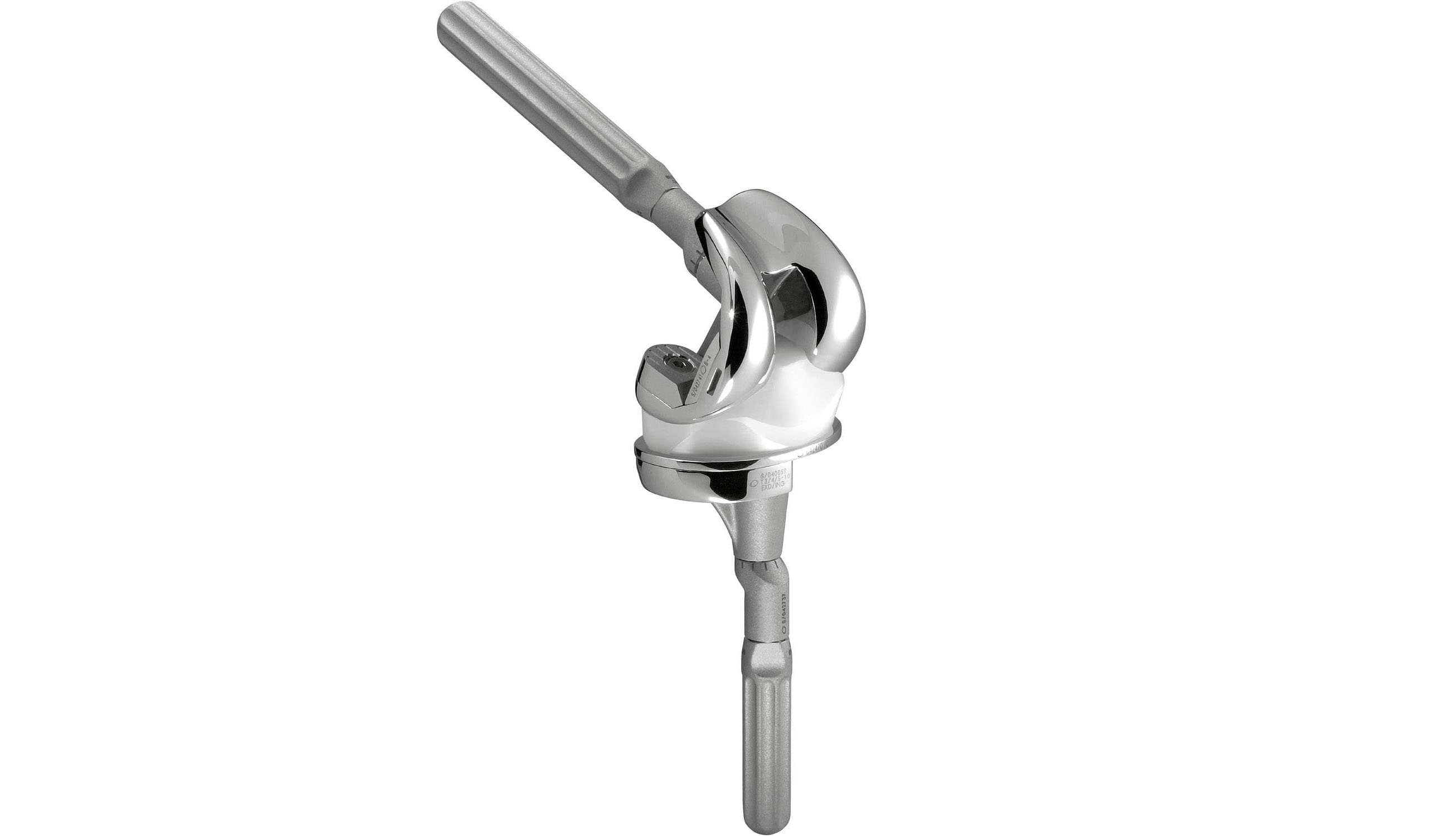
The intervention consists in replacing the damaged joint with a hip prosthesis.
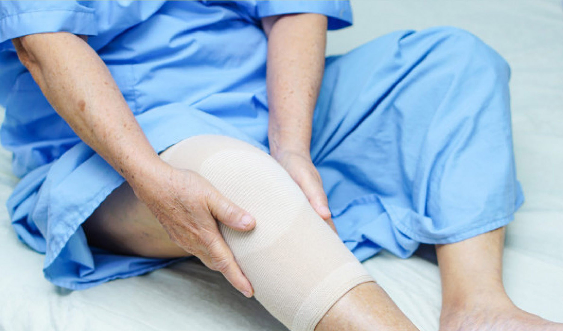
After a rehabilitation program after the intervention, the patients recover and notice the disappearance of the symptoms, being able to walk and realize a practically normal life.
The treatment of the hip prosthesis when there is a problem in this joint requires a great experience in the diagnosis and its realization.
In more than 80% of cases of hip osteoarthritis it is necessary to resort to total replacement of the joint.
The need to replace the hip prosthesis 10 or 15 years after its placement is the main problem. When changing it, it is necessary to put a bigger one and, in each substitution, a smaller adequate bone surface is available to anchor the prosthesis. This is worse in patients under 65, who may need at least two other replacements of this joint throughout their lives.
The placement of a hip prosthesis is an important intervention, but nowadays it is done routinely and with great guarantees in terms of results. ”
Hip osteoarthritis is more common in older people.
If it occurs in a young person, it is usually because the affected hip has suffered some previous illness: congenital dislocation, trauma or some type of inflammation or infection.
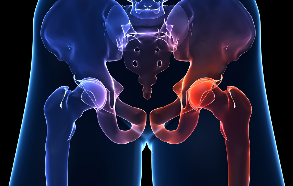
The hip joint is formed by the union between the bones of the pelvis and the femur. In the portion of the femur that joins the pelvis, the femur has the shape of a sphere that is called the head of the femur. This sphere of the femur fits into a hollow that exists in the pelvis, in such a way that a perfect gear is formed that allows the movement of the femur in many directions.
The head of the femur and the hollow of the pelvis in which it articulates are covered with cartilage, which facilitates movements between the bones and prevents them from directly rubbing bone with bone. Over the years and because of the progressive wear of these cartilages, their thickness and texture are lost and can disappear. Thus, the correct gear between the femur and the pelvis is lost, and this is what produces the symptoms of hip osteoarthritis.
Learn more about your health and well-being at Pharmamedic.
[/et_pb_text][/et_pb_column][/et_pb_row][/et_pb_section]